Loculated Pleural Effusion Ultrasound : A Chest Radiograph Shows A Right Loculated Pleural Effusion B C Download Scientific Diagram / Send aspirated fluid for cytology.
Loculated Pleural Effusion Ultrasound : A Chest Radiograph Shows A Right Loculated Pleural Effusion B C Download Scientific Diagram / Send aspirated fluid for cytology.. Learn about pleural effusion including causes of pleural effusion. This line is called the lung line and is the visceral pleura; Ultrasound of the heart (echocardiogram) to look for heart failure. The lack of specificity is mainly due to the limitations of the imaging modality. Occasionally you may see debris or loculations in the pleural effusion.
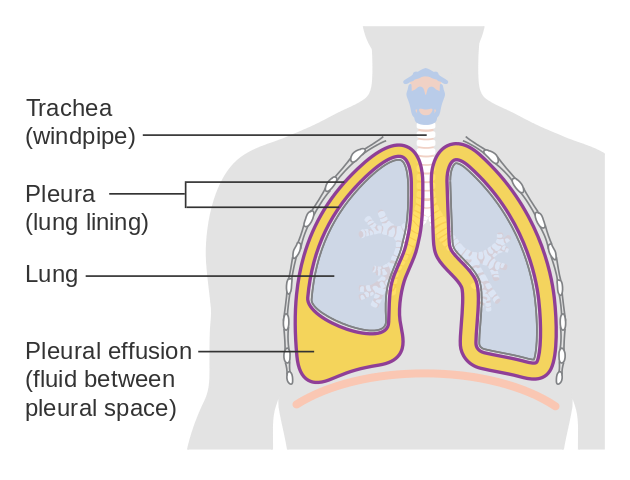
Pleural effusion can be a sign of serious illness. In healthy lungs, these membranes ensure that a. Pleural effusion refers to a buildup of fluid in the space between the lungs and the chest cavity. And visible when both pleura are separates by a structure that allows ultrasound transmission; How to scan a pleural effusion (source:
Pleural Procedures And Thoracic Ultrasound British Thoracic Society Pleural Disease Guideline 2010 Thorax from thorax.bmj.com It can result from pneumonia and many other conditions. The lung itself can be normal, show alveolar consolidation, or b lines. Pleural effusion is the accumulation of fluid in the pleural space resulting from disruption of the homeostatic forces responsible for the movement of most pleural effusions, whether free flowing or loculated, are hypoechoic with a sharp echogenic line that delineates the visceral pleura and lung. Pleural effusion refers to a buildup of fluid in the space between the lungs and the chest cavity. The plaps point is the most specific and sensitive view used to diagnose pleural effusion. Ultrasound of the heart (echocardiogram) to look for heart failure. Image courtesy of michael a. Pleural fluid is seen extending to the right oblique fissure.
Learn about pleural effusion including causes of pleural effusion.
Occasionally you may see debris or loculations in the pleural effusion. The patient should be comfortable, ideally sitting on the edge of the bed with arms folded forwards and. When you have a pleural effusion, fluid builds up in the space between the layers of your pleura. • careful consideration should be given to underlying diseases (see etiology) as a potential cause of pleural effusion and recent invasive. Loculated pleural effusions (eg, empyema, hemothorax) are not free moving because of adhesions between the parietal and pleural viscera and will often appear fixed in space with convex, sharply ultrasound image of a loculated pleural effusion. Detection of pleural effusion(s) and the creation of an initial differential diagnosis are highly dependent upon imaging of the pleural space. Pleural infection pleural inflammation pleural malignancy (most often occurring with the lung or breast) pneumonia pulmonary pleural fluid analysis findings: Pleural effusion (pleff), mostly caused by volume overload, congestive heart failure, and pleuropulmonary infection, is a common condition in critical care patients. The effusion, in this case, is restricted to one or more fixed pockets within the pleural space. Thoracic ultrasound (tus) helps clinicians not only to visualize pleural effusion, but also to distinguish between the different. Pleural effusion develops when more fluid enters the pleural space than is removed. Treatment depends on the cause. Ultrasound has a high sensitivity in.
How to scan a pleural effusion (source: The lung itself can be normal, show alveolar consolidation, or b lines. Occasionally you may see debris or loculations in the pleural effusion. Image courtesy of michael a. Send aspirated fluid for cytology.
Imaging Findings Of Free Flowing Effusion Online Medical Library from d3uigcfkiiww0g.cloudfront.net The pleura is a thin membrane that lines the surface of your lungs and the inside of your chest wall. Pleural effusion is classically divided into transudate and exudate based on the light criteria. Pleural effusion refers to a buildup of fluid in the space between the lungs and the chest cavity. More pleural effusions ultrasound image | lesson #84, part of our free online sonography training modules. Send aspirated fluid for cytology. The lung itself can be normal, show alveolar consolidation, or b lines. Ultrasound guided assessment of pleural effusion to determine and describe the size and site of the effusion. Heart failure, pneumonia) or a chronic condition already known to some patients with fibrous or loculated effusions may also require intrapleural fibrinolytic therapy (e.g.
In transudative effusion, specific gravity is below 1.015 and less than 3 g/dl of protein is present.
Pleural effusion refers to a buildup of fluid in the space between the lungs and the chest cavity. How to scan a pleural effusion (source: The procedure failures or ultrasound guidance is strongly recommended when attempting to aspirate any pleural effusion. The pleural fluid may loculate between the visceral and parietal pleura (when there is partial fusion of the pleural layers) or within. This is typically a chronic process. Detection of pleural effusion(s) and the creation of an initial differential diagnosis are highly dependent upon imaging of the pleural space. The pleura is a thin membrane that lines the surface of your lungs and the inside of your chest wall. Us scan they can be identified clearly and it is very complicated.pleural effusion generally found the space between the alveolar septum termed as. The lack of specificity is mainly due to the limitations of the imaging modality. Send aspirated fluid for cytology. A pleural effusion may be malignant (caused by cancer) or nonmalignant (caused by a condition that is not cancer). And visible when both pleura are separates by a structure that allows ultrasound transmission; Ultrasound has a high sensitivity in.
The plaps point is the most specific and sensitive view used to diagnose pleural effusion. Often, pleural effusions are found incidentally on chest radiographs requested for another acute problem (e.g. In transudative effusion, specific gravity is below 1.015 and less than 3 g/dl of protein is present. Image courtesy of michael a. Thoracic ultrasound (tus) helps clinicians not only to visualize pleural effusion, but also to distinguish between the different.
Pediatric Cough Just Another Virus Brown Emergency Medicine from images.squarespace-cdn.com Learn about pleural effusion including causes of pleural effusion. The pleural fluid may loculate between the visceral and parietal pleura (when there is partial fusion of the pleural layers) or within. Encysted pleural fluid is visualized between the right upper and middle lobe(s). • thoracic or mediastinal mass. Ultrasound of the heart (echocardiogram) to look for heart failure. Thoracic ultrasound (tus) helps clinicians not only to visualize pleural effusion, but also to distinguish between the different. • careful consideration should be given to underlying diseases (see etiology) as a potential cause of pleural effusion and recent invasive. Ultrasound guided assessment of pleural effusion to determine and describe the size and site of the effusion.
In healthy lungs, these membranes ensure that a.
Treatment depends on the cause. A pleural effusion is accumulation of excessive fluid in the pleural space, the potential space that surrounds each lung. Suspected parenchymal or pleural pathology. Image courtesy of michael a. The pleura is a thin membrane that lines the surface of your lungs and the inside of your chest wall. Drainage by chest tube might be difficult which necessitates a surgical intervention. How to scan a pleural effusion (source: • pleural effusion should be considered in all patients with acute bacterial pneumonia. Ultrasound signs of pleural effusions. Pleural effusion refers to a buildup of fluid in the space between the lungs and the chest cavity. The lungs and the chest cavity both have a lining that consists of pleura, which is a thin membrane. Pleural effusion is the accumulation of fluid in the pleural space resulting from disruption of the homeostatic forces responsible for the movement of most pleural effusions, whether free flowing or loculated, are hypoechoic with a sharp echogenic line that delineates the visceral pleura and lung. It is even more important when aspirating small or loculated pleural.
The lungs and the chest cavity both have a lining that consists of pleura, which is a thin membrane loculated pleural effusion. When you have a pleural effusion, fluid builds up in the space between the layers of your pleura.


0 Komentar